
For scientists, precision is everything. From studying the shapes of cells to investigating the functions of proteins, research relies on high-tech tools to advance our understanding of health and disease.
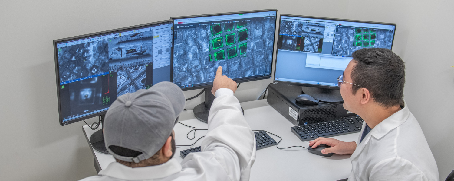
We recently installed one of these powerful technologies at VAI: an Arctis Cryo-Plasma-Focused Ion Beam (cryo-PFIB), a robust sample preparation and microscopy platform that will help us answer some of biology’s most complex questions.
What is cryo-PFIB?
Cryo-PFIB is a leading-edge technology that helps scientists see the innerworkings of cells and tissues in unprecedented detail. It solves a longstanding challenge in biology: some samples are just too thick to see through, and cutting thin slices is a delicate procedure that often ends up damaging or distorting samples.
Cryo-PFIB can cut frozen cells or tissue samples into ultra-thin sections with minimum damage, while keeping the underlying structures intact. The samples are then imaged by high-powered electron microscopes. The result is a transparent cross-section of the sample that shows the exact positions of internal structures and their relationship to one another.
“When an area of interest is so small, it’s hard to know where to cut on the samples,” said Dr. Huilin Li, the Ralph and Grace Hauenstein Endowed Chair in Structural Biology and Chair of VAI’s Department of Structural Biology. “Cryo-PFIB helps us produce more precise results efficiently, while giving scientists a true-to-life look at cells and tissues.”
Learn more about structural biology at VAI ➔
Why is cryo-PFIB important?
Cryo-PFIB helps scientists better understand how cells work in health and how they fail in disease. These insights can inform development of new prevention, diagnosis and treatment strategies.
“VAI is home to world-class technology and the cryo-PFIB is another major step forward for our research,” said Dr. Gongpu Zhao, director of VAI’s Cryo-EM Core. “The cryo-PFIB will enable more precise and efficient sample preparation, allowing us to visualize life’s smallest components in ways we couldn’t explore before.”
In addition to improving study of complex samples, cryo-PFIB provides important insights into cells and molecules in their natural environment, revealing how they behave and interact in context. This approach helps link structure to function, including changes associated with disease states, biological processes or treatment responses.
Fueling discovery at VAI
The new platform already is propelling discovery at VAI.
Assistant Professor Dr. Yang Yang studies how Alzheimer’s and other neurodegenerative diseases begin. She focuses on Alzheimer’s-related plaques, which occur when amyloid-beta proteins in the brain stick together, disrupt cellular communication and contribute to cell death.
“The cryo-PFIB offers me an opportunity to take a closer look at the proteins that comprise these plaques and determine how their structure impacts Alzheimer’s development,” said Yang. “These critical insights could one day lead to improved treatment strategies for neurodegenerative diseases and more.”
Assistant Professor Dr. Travis Walton studies the cellular cytoskeleton, an intricate network of fibers that give cells their shape. Problems with the cytoskeleton are linked to many different diseases, such as Alzheimer’s and Parkinson’s.
“The cytoskeleton is highly specialized for different parts of the body, which means we must view it within the context of tissues,” Walton said. “This new microscope will allow us to see both the molecular details of cytoskeletal function and how it helps organize the inside of cells.”