Scientists now have a powerful new resource for developing better treatments for common cancers of the esophagus, stomach, colon and rectum, collectively known as gastrointestinal (GI) adenocarcinomas. In all, these cancers claim more than 1.4 million lives annually across the globe.
For more than 50 years, GI adenocarcinomas have largely defied therapeutic progress, stymying attempts to develop more effective medications and reduce the necessity of invasive surgeries. Now, thanks to landmark findings from The Cancer Genome Atlas — a joint effort between the National Cancer Institute (NCI), the National Human Genome Research Institute (NHGRI) and hundreds of scientists across six continents — and published this week in the prestigious journal Cancer Cell, researchers have a comprehensive resource for understanding the minute molecular changes that differentiate one type of GI cancer from another.
[This week’s comprehensive analysis of gastrointestinal cancers was published along with 26 other studies detailing similar resources for many other cancer types. Read the announcement here.]

“The better we are able to classify cancers based on their specific molecular characteristics, the better we can treat patients, both by developing new, more targeted therapies and by using this information to inform treatment decisions,” said Dr. Peter W. Laird, a professor at Van Andel Research Institute who was one of the leaders of the GI cancer study. “For GI cancers, our results confirmed some findings from smaller studies that focused on a single cancer type and also revealed differences between and within subtypes that have never before been described, which give us new opportunities to develop improved therapeutic strategies.”
So, what exactly did they find? Why is it important? And what does it mean for the future of cancer research and treatment? Here are the top three takeaways:
TAKEAWAY ONE
GI adenocarcinomas can be divided into at least five subtypes based on molecular characteristics, a departure from traditional classification methods that use anatomic and tissue-specific features to differentiate between cancers.
Historically, cancers have been categorized and named based on the organ or tissue in which they arose — for example, cancers that start in the esophagus have been called esophageal cancers and were believed to be similar to other cancers found in the esophagus, and so on.
The findings urge a shift away from this view, based on new clarity into the incredibly complex web of factors that influence and differentiate one cancer from another.
Rather than categorize by organ of origin, GI cancers should be classified by variations in genetic, epigenetic and molecular makeup, including:
- The presence of Epstein-Barr virus (EBV), a common virus that has been linked to stomach cancer, and correlates to a specific type of epigenetic fingerprint.
- Microsatellite instability (MSI), which is caused by errors in the repair processes responsible for ensuring the genetic code is properly copied. These cancers may be particularly susceptible to immunotherapies.
- Hypermutated tumors with elevated single nucleotide variants (HM-SNV), which have an extreme number of genetic mutations brought on by problems with the polymerase enzyme that copies the DNA during replication. Often, these tumors have a better prognosis than other subtypes due to their increased mutational load, which can cause the body’s immune system to attack or, if a mutation occurs in a gene required for cell viability, can make it difficult for the cancer cell to survive at all.
- Chromosomal instability (CIN), which refers to problems in the 23 pairs of chromosomes that warehouse our DNA.
- Genome stability, or the ability of the genetic code to retain its integrity and pass on an accurate copy of our genetic instruction manual to the next generation of cells.
In short, GI cancers arise in different organs but their similar environment and exposure to the same insults (such as viruses or environmental toxins) mean that these five subtypes transcend organ of origin. A stomach cancer marked by high levels of microsatellite instability, for example, likely has more in common with a colon cancer with the same molecular characteristics than it would with another stomach cancer characterized by Epstein-Barr virus.
TAKEAWAY TWO
More precisely classifying cancers according to molecular makeup has major implications for cancer research and treatment.

When it comes to combating cancer, the old adage, “Know thy enemy” is incredibly apt. Not only do the specific characteristics identified in the findings reveal new vulnerabilities that can be targeted by future medications, but they also may help simplify treatment decisions today. For example, if physicians know that an individual’s cancer has a certain genetic characteristic, they can choose medications designed specifically for that subtype and avoid other treatments that are better suited for a different subtype.
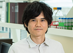
“By identifying the molecular ‘fingerprints’ for each subtype, TCGA has given scientists and physicians an invaluable tool for improving patient care,” said Dr. Toshinori Hinoue, a bioinformatics scientist in Laird’s lab who led much of the analysis for the study. “Even within the five subtypes, we found subtle differences that can be leveraged to inform treatment. It is our hope that this data will lead to new, more effective medications that lessen the need for invasive surgeries and burden on patients’ quality of life.”
TAKEAWAY THREE
Working together is the way forward.
The findings are the result of more than a decade of work by hundreds of scientists around the world who painstakingly analyzed more than 10,000 samples from 33 different cancer types (for the GI study alone, more than 900 samples from esophageal, stomach, colon and rectal cancers were analyzed and compared to nearly 3,000 non-GI cancers).
None of this would have been possible without an extraordinary level of cooperation, teamwork and a singular dedication to creating a resource that could revolutionize cancer research and treatment.
“Team science endeavors like TCGA are the future,” Laird said. “By sharing resources, expertise and data, we were able to do more together than we ever could have apart. The enduring legacy of TCGA will not only be its outstanding contributions to our understanding of cancer, but also serves as proof positive that large-scale collaborations can and do have a major impact.”
All of this week’s TCGA papers can be found at www.cell.com/consortium/pancanceratlas. TCGA data is accessible via NCI’s Genomic Data Commons Data Portal.
Laird is senior author on the paper, along with Adam J. Bass, M.D., of Harvard University and Dana-Farber Cancer Institute, and Vésteinn Thorsson, Ph.D., of the Institute for Systems Biology. Yang Liu, Ph.D., and Nilay S. Sethi, M.D., Ph.D., of the Broad Institute and Dana-Farber Cancer Institute; and Barbara G. Schneider, Ph.D., of Vanderbilt University Medical Center, are co-first authors along with Hinoue.
The manuscripts highlighted in this release are part of The Cancer Genome Atlas (TCGA) Program, a joint effort of the NationalCancer Institute (NCI) and the National Human Genome Research Institute (NHGRI).
Research reported in this publication was supported by the Intramural Research Program and the following grants from the National Institutes of Health: U24 CA143799, U24 CA143835, U24 CA143840, U24 CA143843, U24 CA143845, U24 CA143848, U24 CA143858, U24 CA143866, U24 CA143867, U24 CA143882 (Laird and Stephen Baylin, Ph.D., Johns Hopkins University/VARI), U24 CA143883, U24 CA144025, U54 HG003067, U54 HG003079, U54 HG003273 and P30 CA16672. The content is solely the responsibility of the authors and does not necessarily represent the official views of the National Institutes of Health.
