Van Andel Research Institute installs world-class microscopes to enable discovery of the molecular basis of disease
March 27, 2017
GRAND RAPIDS, Mich. (March 28, 2017)—Van Andel Research Institute (VARI) is now home to one of the world’s most powerful microscopes—one that images life’s building blocks in startling clarity and equips VARI’s growing team of scientists to push the limits of discovery in search of new treatments for diseases such as cancer and Parkinson’s.
Cryo-electron microscopy (cryo-EM) is a revolutionary technique that reveals in unprecedented detail the molecular and atomic interactions at the foundation of life, allowing scientists to observe exactly how viruses enter cells, how DNA replicates, how chemical compounds assemble into functional biological components, and much more.
“Our new state-of-the-art cryo-EM facility, which includes significant investments in technology and talent, is part of our unwavering commitment to improve human health through scientific innovation,” says David Van Andel, the Institute’s CEO and chairman. “Not only will it fuel the discovery of life-changing treatments for devastating diseases, but it also will enhance Grand Rapids’ reputation as a destination for outstanding biomedical research.”
Already, cryo-EM has helped uncover the complex structure of Alzheimer’s-related proteins in the brain, overturned long-held assumptions about DNA replication and elucidated the structure of hundreds more molecules implicated in a variety of diseases.
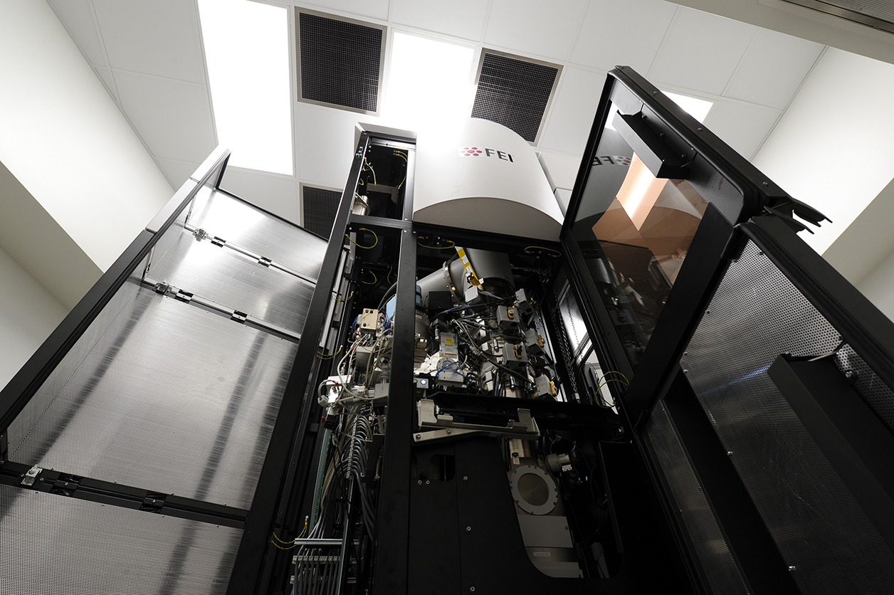
The centerpiece of VARI’s cryo-EM facility is an FEI Titan Krios from Thermo Fisher Scientific, the world’s highest-resolution, commercially available cryo-EM. The Institute’s Krios is only the second in Michigan and one of fewer than 100 in the world. The facility also houses an FEI Talos Artica and an FEI Tecnai Spirit G2 BioTWIN. Overall, VARI invested $10 million in cryo-EM equipment and related construction.
“The resolution available with the Krios, compared to earlier methods, is akin to upgrading from a road atlas to Google Earth,” says Peter Jones, Ph.D., D.Sc., VARI’s chief scientific officer. “It offers exquisite detail of complex systems, which will help us find new therapies so desperately needed for patients around the world.”
Part of the expansion also included hiring three cryo-EM experts: Huilin Li, Ph.D., an established investigator in the field with more than 20 years of experience; Wei Lü, Ph.D., an early career investigator who joined the Institute this month; and Gongpu Zhao, Ph.D., the cryo-EM facility’s manager, who played a key role in the 2013 discovery that revealed the structure of the HIV-1 virus’s outer shell.
TECHNOLOGICAL BREAKTHROUGHS
Although cryo-EM has been around for decades, recent advances in both techniques and technology have revolutionized the approach, giving researchers powerful new tools to more quickly and more precisely see some of the smallest yet most important biological components in their natural state. These advances led to it being named as the Method of the Year in 2015 by the scientific journal Nature Methods.
“We are very pleased that VARI has chosen the Titan Krios. We have worked hard with leading life science researchers to perfect the capabilities they need to advance their understanding of living systems and disease processes,” says Peter Fruhstorfer, vice president and general manager of Life Sciences, Materials and Structural Analysis, at Thermo Fisher. “The advanced imaging capabilities of the Titan Krios allows scientists to visualize delicate biological structures at the molecular level without damage and in a nearly-natural context, helping them understand the essential relationships between structure and function that are the basis for life itself.”
Before the advances in cryo-EM, a technique called X-ray crystallography was the dominant method of determining molecular structure. However, the approach is very slow and painfully difficult because biological molecules are hard to crystallize. In cryo-EM, the need for crystallization is entirely circumvented. Scientists simply flash freeze molecules or cells in solution. The molecules, now embedded in a transparent thin layer of glassy ice, are then scanned with a powerful electron beam, generating hundreds of thousands of two-dimensional images that are then assembled in a computer into a detailed three-dimensional portrait.
“In a way, biologists are like locksmiths,” says Li. “We use tools such as cryo-EM or X-ray crystallography to see all of the facets of the lock— a normal protein doing its job in a cell or an abnormal protein in cancer, for example—and then use that information to design a key to fit the lock—a drug with the right shape to link up with the protein and alter its function, which may correct the error, ultimately treating the disease.”
###
ABOUT VAN ANDEL RESEARCH INSTITUTE
Van Andel Institute (VAI) is an independent nonprofit biomedical research and science education organization committed to improving the health and enhancing the lives of current and future generations. Established by Jay and Betty Van Andel in 1996 in Grand Rapids, Michigan, VAI has grown into a premier research and educational institution that supports the work of more than 360 scientists, educators and staff. Van Andel Research Institute (VARI), VAI’s research division, is dedicated to determining the epigenetic, genetic, molecular and cellular origins of cancer, Parkinson’s and other diseases and translating those findings into effective therapies. The Institute’s scientists work in onsite laboratories and participate in collaborative partnerships that span the globe. Learn more about Van Andel Institute or donate by visiting www.vai.org. 100% To Research, Discovery & Hope®
Huilin Li, Ph.D.
Cryo-EM Core Director
Professor, Center for Epigenetics
Wei Lü, Ph.D.
Assistant Professor, Center for Cancer and Cell Biology
Gongpu Zhao, Ph.D.
Cyro-EM Core Manager
Read our blog post “Why we’re betting on cryo-electron microscopy” by clicking here.