Why we’re betting on cryo-electron microscopy
March 29, 2017
We just invested in a new Cryo-Electron Microscope (Cryo-EM) Core facility, including installation of one of the world’s most powerful microscopes and recruitment of several outstanding scientists. Here’s why.
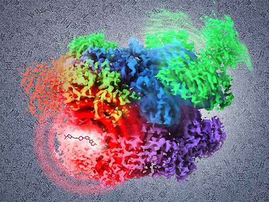
The imaging resolution available with the FEI Titan Krios from Thermo Fisher Scientific, the largest of our microscopes, is akin to watching a bouncing tennis ball on Earth from the surface of the moon. The possibilities with that kind of visual power are immeasurable. We believe cryo-EM has the potential to usher in a wave of discovery that parallels a handful of crucial developments in the history of science.
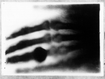
Consider the X-ray. Invented in 1895 by German physicist Wilhelm Röntgen, the first X-ray machine was bulky, expensive and technically difficult to build. However, early adopters of these machines, delivered groundbreaking and prolific contributions to our understanding of the human body.
Suddenly, scientists could see kidney stones passing into the ureter, phalanges of hand deformities, food moving through the digestive system, and a penny lodged in a child’s throat.
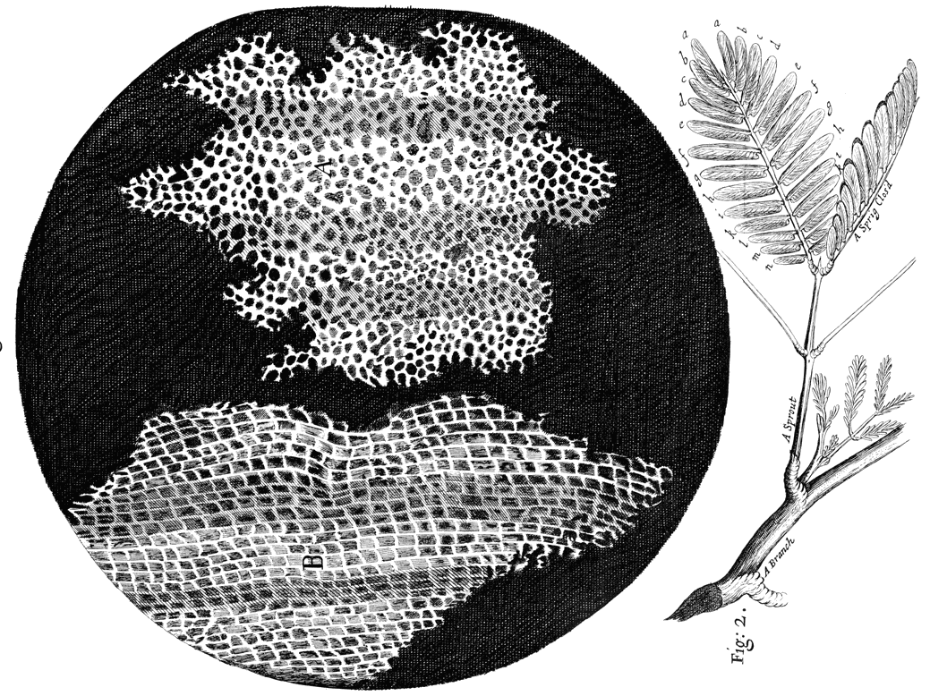
Similarly, the first compound microscopes led to discovery of several fundamental tenets of biology. Dutch scientists discovered cells by observing the honeycomb structure of a sliver of cork. They offered visual confirmation of bacteria, which were teeming in a droplet of lake water. They revealed the stunning complexity of tiny insects like fleas, lice and gnats.
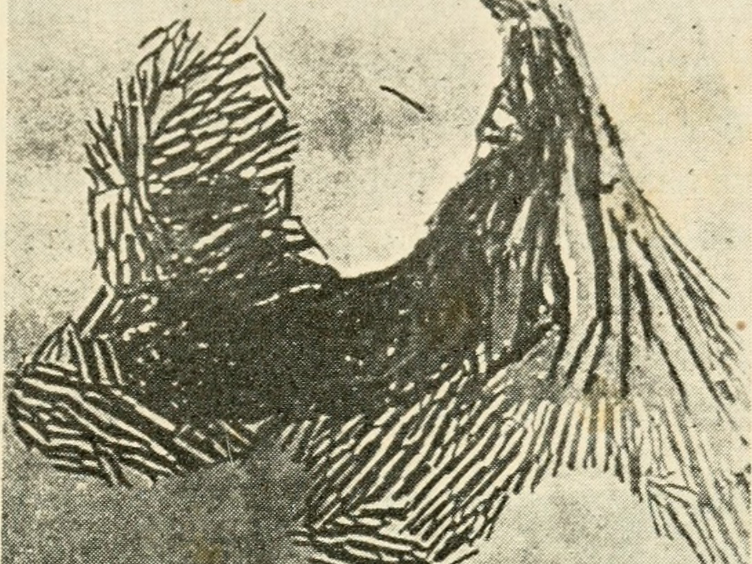
Analogous breakthroughs in imaging abound. The first telescopes offered irrefutable visuals of the sun as center of our solar system. Electron microscopes allowed for the first images of DNA and viruses.
Now, the latest generation of cryo-electron microscopes are revealing the atomic and molecular interactions at the foundation of life. Scientists can see, for the first time, the exact size, shape, and function of profoundly complex proteins and enzymes.
We can watch as chemicals inside a nucleus assemble and begin the process of DNA replication. We can visualize the atom-by-atom structure of α-synuclein proteins, which are thought to impair the brains of people with Parkinson’s disease. We can image cell-signaling pathways thought to encourage tumor growth. The list goes on.
We believe these discoveries are only the beginning. And we believe cryo-EM represents a critical component of our unwavering commitment to improving the health and enhancing the lives of current and future generations.